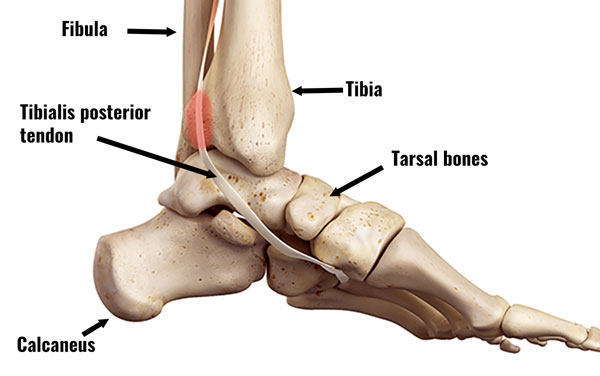
what bones does the posterior tibial tendon attach to
Tibialis posterior tendonitis (tendinopathy) is an overuse injury causing pain on the inside of the talocrural joint. Pain is felt on the inside of the ankle which may radiate nether the curvation of the pes. Here nosotros explicate the symptoms, causes and treatment of Tibialis posterior tendonitis.
Tibialis posterior tendinopathy symptoms
Symptoms include:
- Hurting on the inside of the ankle, specifically backside the medial malleolus. This is the bony protrusion on the inside of the ankle.
- Hurting comes on gradually over time.
- Symptoms may besides radiate along the length of the tendon as it passes under the human foot.
- A creaking sensation called crepitus during movement.
In the early stages swelling is unlikely. Just later you may encounter local swelling around the back of the malleolus. If you take a lot of swelling, then you may have an acute partial tear of the tendon.
Diagnosis & cess
Your physio or doctor will practice a number of tests to assess your ankle.
Passive eversion
This is simply stretching your tibialis posterior whilst you stay relaxed. Your therapist turns the foot outwards. If this is painful and so the test is positive.
Resisted inversion
This is when the patient turns the soles of the anxiety inwards whilst the therapist resists the motion. If pain is produced then the exam is positive.
Heel enhance
Some other exam is to view the athlete from backside performing a heel raise on a step. If they have posterior tibialis tendinopathy then they may have difficulty keeping the talocrural joint straight.
It volition remain rolled in and flattened. In astringent cases the patient volition take difficulty doing a heel raise altogether.
Avulsion strains
If there is pregnant hurting under the foot and then this may betoken a partial avulsion where the tendon pulls away from the bone at the attachment to the navicular os.
Imaging
MRI – an MRI or magnetic resonence imaging can confirm the diagnosisane.
What is Tibialis posterior tendonitis?
Tibialis posterior tendonitis is an overuse injury causing inflammation (or degeneration) of the Tibialis posterior tendon.
Tendinopathy is probably a more accurate term. This is because it refers to wear and tear or degeneration of the tendon. Long term, chronic injuries are more likely to be wear and tear because acute inflammatory cells are not usually nowadays.
The tibialis posterior muscle passes down the dorsum of the leg and under the medial malleolus. This is the bony chip on the within of the ankle. Information technology inserts into the post-obit basic in the pes:

- Navicular and cuneiform bones in the midfoot.
- The base of the 2nd, 3rd, fourth and fifth long metatarsal basic under the foot.
It plantarflexes the foot (going up on your toes) and inverts the pes (turning the soles of the feet inwards).
Causes
Athletes who overpronate are at increased risk. This is considering there is more strain on the tibialis posterior muscle. Equally a result, it repeatedly overstretches and overworks.
It is near likely to bear upon older female athletes, especially those who walk or run.
Sports and activities that increase the take a chance of tibialis posterior tendinopathy are those requiring prolonged stretching of the tibialis posterior muscle. For case:
- Ballet dancing
- Water ice skating
- Sprinters who run a lot on tight bends
Long-term injuries to the tibialis posterior result in insufficiency of the musculus and a condition called tibialis posterior dysfunction (PTTD) which results in fallen arches, or apartment feet.
At that place is sometimes confusion between PTTD and tibialis posterior tendinopathy and the terms are often used interchangeably. However, these conditions are slightly different as PTTD is a dysfunction of the muscle and tendinopathy is a degeneration of the tendon.
Treatment for Posterior tibial tendonitis
Immediate start aid is to use the Toll principles of protection, rest, water ice, compression, and elevation. And so a full ankle rehabilitation program gets you back to full fitness.
Cold therapy
Utilise ice or cold therapy for 10 minutes every 60 minutes for the outset 24 to 48 hours, especially if painful, tender or inflamed. Reduce frequency to 3 times per day equally your symptoms improve.
Do not apply icedirectly to the peel just wrap in a wet tea towel. Or employ a commercially available common cold compression wrap.
Afterwards, after the acute stage, estrus may be more benign.
Compression
Utilize a tube grip bandage, ankle support or taping. This protects your ankle and reduces swelling.
Corticosteroid injections should NOT be used to care for tibialis posterior tendinopathy. It increases the risk of a complete tendon rupture.
Electrotherapy
Your physio may apply electrotherapy for example, ultrasound to help with pain and swelling.
Medication
A doc may prescribe anti-inflammatory medication for case Ibuprofen. Short-term this reduces inflammation. All the same, less likely to help long term. Always check with a dr. before taking medication and practise not take ibuprofen if you take asthma.
Massage
Apply cross friction sports massage techniques to the tendon and deep tissue massage to the tibialis posterior and calf muscles. This may help increment flexibility and condition of the muscles.
Orthotics
Orthotic shoe inserts correct poor foot biomechanics.
Surgery
If your tendon has ruptured completely, then it needs surgery to repair it.
Exercises
When hurting allows, brainstorm stretching exercises for the tibialis posterior and calf muscles. Specific exercises for the tibialis posterior strengthen the muscle. As a event, this helps preclude future injury.
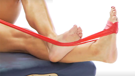
Begin strengthening exercises every bit soon as they are pain costless. In particular, eccentric strengthening for the tibialis posterior. This ways strengthening the muscle at the same time as it lengthens.
Read more than on Tibialis posterior strengthening exercises.
References
- Chhabra A, Soldatos T, Chalian G, three-tesla magnetic resonance imaging evaluation of posterior tibial tendon dysfunction with relevance to clinical staging. J Foot Ankle Surg 2011;fifty:320–328
hughes-jonesroadvine1969.blogspot.com
Source: https://www.sportsinjuryclinic.net/sport-injuries/ankle-pain/medial-ankle-pain/tibialis-posterior-tendinopathy
0 Response to "what bones does the posterior tibial tendon attach to"
Post a Comment